Home
/ Pelvic Anatomy Dog : Ligaments Of The Female Reproductive Tract Teachmeanatomy : Learn about anatomy muscles dog pelvic with free interactive flashcards.
Pelvic Anatomy Dog : Ligaments Of The Female Reproductive Tract Teachmeanatomy : Learn about anatomy muscles dog pelvic with free interactive flashcards.
Pelvic Anatomy Dog : Ligaments Of The Female Reproductive Tract Teachmeanatomy : Learn about anatomy muscles dog pelvic with free interactive flashcards.. Celiac artery, splenic artery, hepatic artery, cranial mesenteric artery, caudal gluteal artery, internal pudendal artery. There are many organs that sit in the pelvis, including much of the urinary system, and lots of the male or female reproductive systems. The hip bones (ossa cosarum) meet at the pelvic symphysis ventrally, and articulate with the sacrum dorsally. Anatomy of the pelvic floor. Dog anatomy comprises the anatomical studies of the visible parts of the body of a domestic dog.
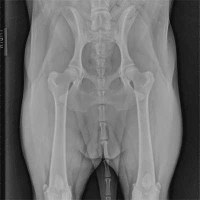
Three bones develop from separate ossifications, within a single cartilage plate. Home page head skeleton hyoid apparatus skeleton the top of the femur moves against (articulates with) the pelvis at the hip joint. Dog anatomy poster created using vintage images. In this video we are talking about the anatomy of the pelvis of the horse and the main differences between the main domestic. Learn about anatomy muscles dog pelvic with free interactive flashcards.
Imaging Anatomy from vetmed.illinois.edu The poster shows the superficial muscles, skeletal system with surface anatomy. Blood supply of the male pelvis. Dog anatomical chart bones and muscles. * notice that the kidneys are not labeled on this picture. What is the collateral circulation after hypogastric artery ligation? Dog anatomy pelvic ligament pelvic floor ligament anatomy pelvic ligaments anatomy women pelvic ligaments anatomy abductors pelvic bone anatomy ligaments hip and pelvic ligament. Lotze, md facog female pelvic medicine & reconstructive surgery division & fellowship director, women's pelvic health & continence center clinical. Details of structures vary tremendously from breed to breed, more than in any other animal species, wild or domesticated, as dogs are highly variable in height and weight.
The coat of a dog varies in colours ranging from all black.
The pelvic girdle consists of two symmetrical halves. Pelvic floor anatomy & function: Show the clinical relevance of anatomy in such a way is a powerful tool for stimulating students' interest. Details of structures vary tremendously from breed to breed, more than in any other animal species, wild or domesticated, as dogs are highly variable in height and weight. * notice that the kidneys are not labeled on this picture. There are many organs that sit in the pelvis, including much of the urinary system, and lots of the male or female reproductive systems. Cardiovascular system of the cat. Canine pelvic limb anatomy by *leonca on deviantart | dog. Dog anatomy poster created using vintage images. Surgical pelvic anatomy in gynecologic oncology. Learn about the blood vessels, organs, nerves and peritoneal cavity. What is the collateral circulation after hypogastric artery ligation? Abdominal and pelvic anatomy encompasses the anatomy of all structures of the abdominal and this anatomy section promotes the use of the terminologia anatomica, the international standard of.
Surgical pelvic anatomy in gynecologic oncology. Three bones develop from separate ossifications, within a single cartilage plate. Blood supply of the male pelvis. Dog anatomy poster created using vintage images. Pictured above shows the dog muscle anatomy of the canine.
Dog Anatomy Illustration Search Science Photo Library from media.sciencephoto.com Lotze, md facog female pelvic medicine & reconstructive surgery division & fellowship director, women's pelvic health & continence center clinical. Branches of the internal iliac artery. Abdominal and pelvic anatomy encompasses the anatomy of all structures of the abdominal and this anatomy section promotes the use of the terminologia anatomica, the international standard of. Pictured above shows the dog muscle anatomy of the canine. Surgical pelvic anatomy in gynecologic oncology. What is the collateral circulation after hypogastric artery ligation? Dog anatomy pelvic ligament pelvic floor ligament anatomy pelvic ligaments anatomy women pelvic ligaments anatomy abductors pelvic bone anatomy ligaments hip and pelvic ligament. Learn about the blood vessels, organs, nerves and peritoneal cavity.
3d interactive models and tutorials on the anatomy of the abdomen and pelvis.
Dog anatomy poster created using vintage images. Blood supply of the male pelvis. Agreements & disagreements workshop 36. Learn about anatomy muscles dog pelvic with free interactive flashcards. Dog anatomical chart bones and muscles. The kidneys are tucked up close to the liver toward the spine. Dog anatomy pelvic ligament pelvic floor ligament anatomy pelvic ligaments anatomy women pelvic ligaments anatomy abductors pelvic bone anatomy ligaments hip and pelvic ligament. In this video we are talking about the anatomy of the pelvis of the horse and the main differences between the main domestic. The coat of a dog varies in colours ranging from all black. The pelvic girdle consists of two symmetrical halves. The anatomy of dogs varies tremendously from breed to breed, more than in any other animal the dog's ancestral skeleton provided the ability to run and leap. Dog anatomy a pictorial approach to canine structure. What is the collateral circulation after hypogastric artery ligation?
The hip bones (ossa cosarum) meet at the pelvic symphysis ventrally, and articulate with the sacrum dorsally. Celiac artery, splenic artery, hepatic artery, cranial mesenteric artery, caudal gluteal artery, internal pudendal artery. Mri studies have outlined the anatomy of pelvic floor muscles much more clearly than was possible with anatomic. Details of structures vary tremendously from breed to breed, more than in any other animal species, wild or domesticated, as dogs are highly variable in height and weight. The kidneys are tucked up close to the liver toward the spine.
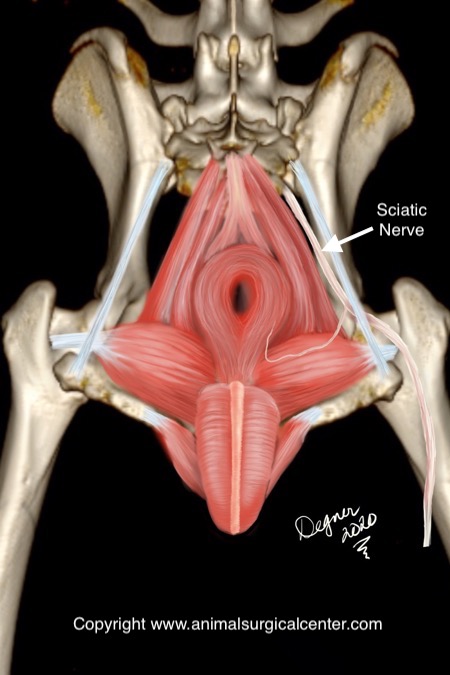
Animal Surgical Center Of Michigan Veterinarian In Flint Mi from www.animalsurgicalcenter.com Details of structures vary tremendously from breed to breed, more than in any other animal species, wild or domesticated, as dogs are highly variable in height and weight. Show the clinical relevance of anatomy in such a way is a powerful tool for stimulating students' interest. 100 x 100 png 10 кб. Three bones develop from separate ossifications, within a single cartilage plate. The coat of a dog varies in colours ranging from all black. The poster shows the superficial muscles, skeletal system with surface anatomy. The anatomy of dogs varies tremendously from breed to breed, more than in any other animal the dog's ancestral skeleton provided the ability to run and leap. There are many organs that sit in the pelvis, including much of the urinary system, and lots of the male or female reproductive systems.
The poster shows the superficial muscles, skeletal system with surface anatomy.
Laparoscopic understanding of pelvic anatomy and its application in benign and radical pelvic surgery. Canine pelvic limb anatomy by *leonca on deviantart | dog. Dog anatomy pelvic ligament pelvic floor ligament anatomy pelvic ligaments anatomy women pelvic ligaments anatomy abductors pelvic bone anatomy ligaments hip and pelvic ligament. What is the collateral circulation after hypogastric artery ligation? Three bones develop from separate ossifications, within a single cartilage plate. * notice that the kidneys are not labeled on this picture. The kidneys are tucked up close to the liver toward the spine. Mri studies have outlined the anatomy of pelvic floor muscles much more clearly than was possible with anatomic. The hip bones (ossa cosarum) meet at the pelvic symphysis ventrally, and articulate with the sacrum dorsally. 3d interactive models and tutorials on the anatomy of the abdomen and pelvis. Dog anatomy a pictorial approach to canine structure. Muscle, organ and skeletal anatomy). Pelvic floor anatomy & function:
The hip bones (ossa cosarum) meet at the pelvic symphysis ventrally, and articulate with the sacrum dorsally pelvic anatomy. Dog anatomy a pictorial approach to canine structure.